Leaving partial dentures in the mouth for a panoramic film will usually obscure important diagnostic information as seen in the above film.
Floor of the orbit panoramic.
Usually only the inferior border of the orbit is visible over the panoramic radiograph.
The orbit is a pear shaped cavity with an apex directed posteriorly medially and slightly upward.
The anatomical landmarks of the are the floor of the orbit and the external auditory meatus.
Styloid process a long pointed radiopaque structure that extends from the temporal bone anterior to the mastoid process bilateral.
On a panoramic radiograph the incisive foramen appears as a small ovoid or round area located.
Orbit a radiolucent area superior to the maxillary sinus bilateral.
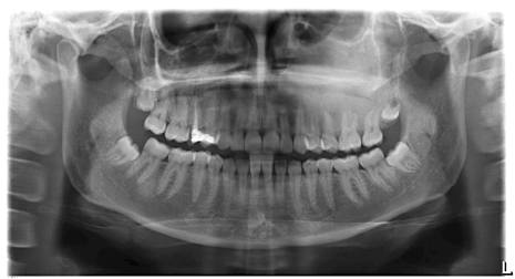
The chin is too far up notice how wide the condyles are and the arc to the hard palate.
Both statements are true.
The orbit is a conical structure with its base facing anterolaterally and its apex originating medially as the inlet of all vital neural and vascular structures via the optic foramen superior orbital fissure and inferior orbital fissure.
Regarding inflammatory conditions of non odontogenic origin these are usually clearly demonstrated on panoramic radiography if they involve mucosal thickenings arising from the floor of the maxillary sinus.
External auditory meatus external acoustic meatus.
It is also bound by the medial and lateral walls.
Preparation to take the panoramic image the clinician needs to have the patient remove jewelry bobby pins hearing aids etc.
A bony cavity containing the eyeball is a radiolucent compartment with radiopaque borders situated superior to the maxillary sinus.
Panoramic radiography when large œ and within the panoramic image layer.
Anatomical landmarks on panoramic radiography enumerate all radiolucent landmarks visible on a panoramic radiograph bony landmarks of the maxilla and surrounding structures.
The forms the floor of the orbit of the eyes the sides and floor of the nasal cavity and the hard palate.
Note the hearing aid in the left ear green arrow and its ghost image overlying the right orbit red arrows.
The anterior rim of the bony orbit the orbital rim is formed by orbital processes from the maxilla z.
The orbit and zygomatic arch.
On a panoramic radiograph the appears as a rounded radiopaque projection of the bone located anterior to the glenoid fossa.
The most frequent example of such a process is the mucous.
Only the border of the orbit is visible on most panoramic radiographs.
The forms the floor of the orbit of the eyes the sides and floor of the nasal cavity and the hard palate.