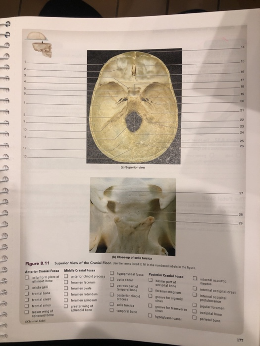
Figure 1 2 saddle like depression which is sella turcica.
Floor of sella turcica bone.
The sella turcica latin for turkish seat is a saddle shaped depression in the body of the sphenoid bone of the human skull and of the skulls of other hominids including chimpanzees orangutans and gorillas it serves as a cephalometric landmark the pituitary gland or hypophysis is located within the most inferior aspect of the sella turcica the hypophyseal fossa.
The sella turcica is a cup shaped depression in the central basisphenoid bone which contains the pituitary gland and inferior part of the infundibular stalk.
Sella tur cica a depression on the upper surface of the sphenoid bone lodging the pituitary gland.
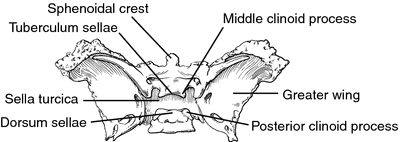
The sellar floor is continuous anteriorly with the tuberculum sellae and posteriorly with the dor sum sellae.
It is divided into the following parts.
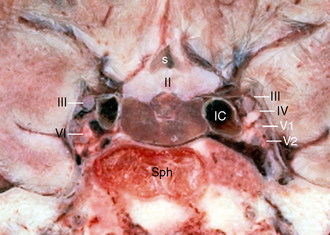
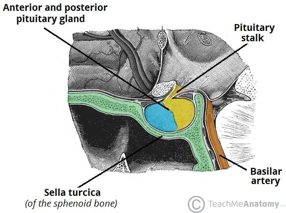
The sphenoid sinus is inferior and anterior to the sella turcica the paired cavernous sinuses are lateral the suprasellar cistern and its contents are superior and the basilar artery and brainstem are posterior the pituitary gland which weighs about 0 5 g in the adult is the only structure of.
Empty sella see empty sella syndrome.
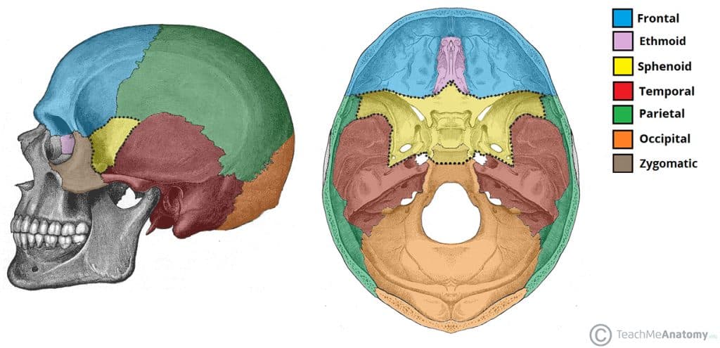
A median portion known as the body of sphenoid bone containing the sella turcica which houses the pituitary gland as well as the paired paranasal sinuses the sphenoidal sinuses.
The sphenoid bone contains two cavities known as the sphenoidal sinuses.
It houses the pituitary gland.
Turcica turkish is a bony saddle shaped structure on the superior surface of the body of the sphenoid figure 1.
The sphenoid bone.
Two greater wings on the lateral side of the body and two lesser wings from the anterior side.
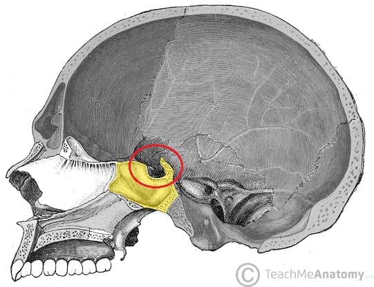
The sella turcica is a depression in the central region of the sphenoid bone.
Figure 14 4 gross anatomy of the sella and pituitary gland.
The anterior part of the sella turcica which forms the horn of the saddle is a ridge called the tuberculum sellae.
Sella sel ah l a saddle shaped depression.
2 not surprisingly many patients.
The sella turcica sella saddle.
Miller keane encyclopedia and dictionary of medicine nursing and.
Coronal cryomicrotome section through the sella turcica.
Sella turcica resembles as saddle like depression which provide place for the pituitary gland.
Pterygoid processes of the sphenoides directed downwards from the junction of.
Anatomically the sella turcica has been expressed as variable.
The sphenoid bone is the large bat shaped bone that spans the floor of the skull.
1 autopsy studies confirm the high disease prevalence reported to be 5 5 to 20 of the general population.
Which of the following bones do not contain a sinus.
It is divided into three fragments and consists of an anterior wall a floor and a posterior wall.
Sella turcica in the superior aspect of the sphenoid bone.
The purplish pituitary gland unlabeled rests on the floor of the sella between the paired cavernous segments of the internal carotid arteries ic.